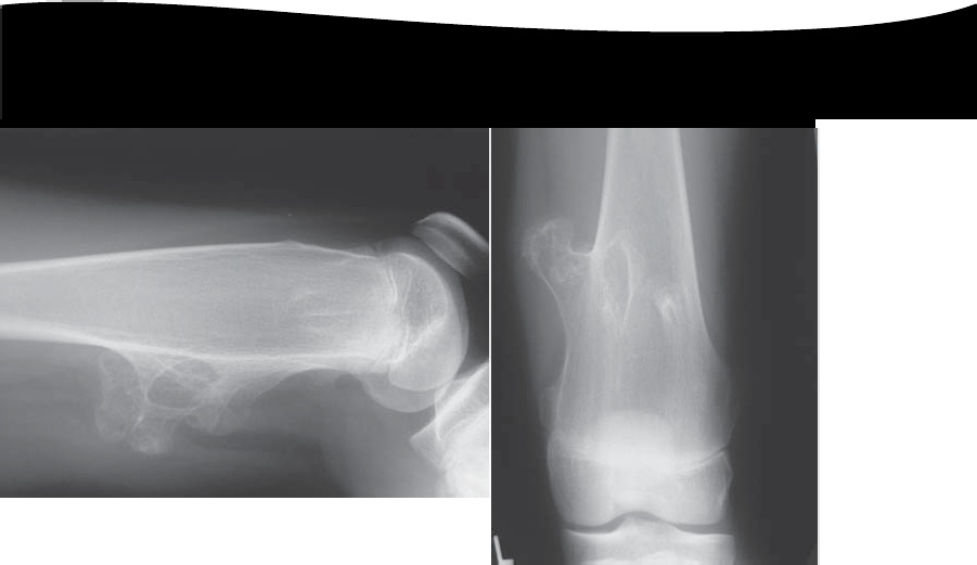
These AP and lateral radiographs show sessile lesions arising from the metaphyseal region of the distal femur.
The lesions are well defined and appear to be growing away from the metaphysis.
The matrix of the lesions is in continuity with the surrounding normal bone.
The cortex of the normal bone appears to be in continuity with the lesions.
The caps of the lesions contain specks of calcification.
These appearances would be compatible with a slow-growing, benign lesion, most likely osteochondroma.
————————————————————————————————————–
Hereditary multiple exostoses (HME) is a familial inherited autosomal dominant condition but spontaneous mutation also occurs.
Males are more often affected, possibly due to an incomplete penetrance in females. Three gene mutations have been identified that can lead to HME.
HME Type I is caused by a mutation in the gene encoding exostosin (EXT1) which maps to chromosome 8q24.
HME Type II is caused by mutation in the gene encoding exostosin-2 (EXT2)
on chromosome 11, and
HME TypeIII has been mapped to a locus on chromosome 19 (EXT3)
The radiographic distribution of lesions is as follows:
Distal femur 70%
Ribs 40%
Proximal tibia 70%
Distal radius 30%
Proximal humerus 50%
Distal ulna 30%
Scapula 40%
Generally, patients present with a painless mass.
The developing exostoses may lead to abnormalities in osseous growth, joint restriction, joint deformities particularly affecting paired bones, and early progression to osteoarthritis.
Lesions that continue to enlarge after the end of puberty are abnormal and should be investigated for potential malignant change (chondrosarcoma).
Ultrasonography and MRI are usually the investigations of choice.
Features suggesting malignant change include:
Increasing pain and swelling (especially after cessation of normal growth)
Thickening of the cartilage cap (>2 cm is very concerning)
Lysis of a proportion of the stalk
Intramedullary invasion of the underlying bone
Leave a Reply