Teaching Points :
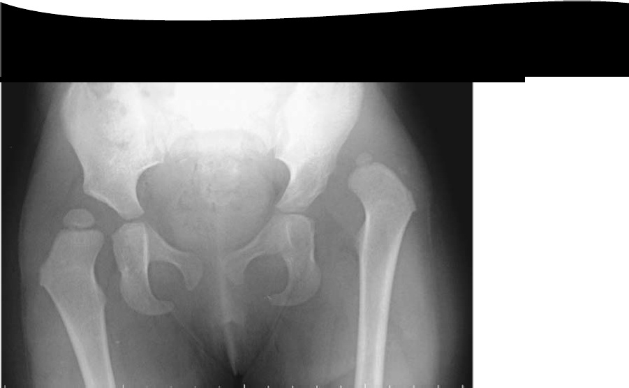
— > This is an AP pelvic radiograph showing a dislocated left hip and dysplastic acetabulum.
— Shenton’s line is broken and the femoral head lies lateral and superior to the inferiomedial quadrant (made by the intersection of Perkin’s and Hilgenreiner’s lines).
–> How would you proceed in you management from here?
** I would take a full history and examine the child.
There may be risk factors for developmental dysplasia of the hip (DDH) including positive family history and/or decreased intrauterine space ,first born,breech, oligohydramnios (associated packaging problems).
**More importantly I would be looking to see if there were any underlying neuromuscular conditions such as spina bifida, arthrogryphosis, or cerebral palsy.
**Examination may reveal a Trendelenberg gait,leg length discrepancy, fixed flexion deformity as well as reduced abduction of the left hip, which is the most consistent and reliable clinical sign of this condition.
** I would organize an examination under anaesthesia (EUA) and arthrogram to delineate the anatomyof the acetabulum, soft tissues, and proximal femur.
** It would be unlikely that this hip would reduce closed.
** Blocks to reduction would include: an inverted limbus; elongated ligamentum teres; hour-glassconstriction of the capsule; psoas tendon and pulvinar.
** Indications for open reduction include: failureof closed reduction; an unstable reducible hip, or soft tissue interposition preventing a congruent reduction.
–> What open operative approaches would you use to reduce this hip?
I would use a modified anterior (ilio-femoral) approach to the hip.
I would place my skin incisionparallel and distal to the iliac crest, passing 2 cm distal to the anterior superior iliac spine (ASIS) and extending medially within the groin skin crease.
I would identify and protect the lateral cutaneous nerve of the thigh and then distally I would develop the internervous plane between tensor fascia lata (superior gluteal nerve) and sartorius (femoral nerve).

Leave a Reply